
TEMP/TDM
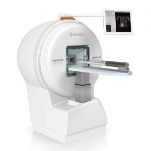
Haute sensibilité, haute résolution, le nanoScan SPECT/CT (TEMP/TDM Tomographie par Emission MonoPhotonique couplée à la TomoDensitoMétrie) est un système intégré pour l’imagerie d’un large éventail d’espèces animales allant des souris à des singes.
C’est un appareil unique et breveté multimodalité qui permet l’imagerie fonctionnelle 3D des traceurs SPECT de la résolution de niveau sub millimétrique . Le micro-CT intégré, ultra-rapide, à faible dose, avec un champ de vue variable fournit des informations anatomiques automatiquement enregistrées pour la fusion et la correction des données.
C’est un appareil haute résolution qui permet l’imagerie du corps entier de petits animaux (souris, rats, furets, cochons nains, des lapins, et même des petits primates non humains).
Résumé des avantages clés pour SPECT
- Ultra Haute Performance (UHP) Détecteur
- Mediso multifocales multi-taille multiples ouvertures pinhole (M3-pinhole™)
- Ouvertures rectangulaires d’une taille d’ouverture variable
- Géométrie multifocale
- Ouvertures appropriées selon l’application : souris corps entier, rat corps entier, ultra-haute sensibilité
- Remplacement rapide et facile des ouvertures
Caractéristiques du sous-système CT
Le Tomographe à rayons X est un système d’imagerie préclinique corps entier.
Permet d’imager la plus grande variété d’espèces dans l’industrie, y compris les grands animaux ou un corps entier de souris du corps en une position allongée. Doté d’une très grande surface de détection et d’un grand accès avec la possibilité de balayage hélicoïdale, un zoom variable (jusqu’à 8) et une source de rayons X haute puissance (jusqu’à 80W).
- Possède la plus large gamme d’application CT: ultrarapide et ultra faible dose d’irradiation avec zoom variable jusqu’à 7.6x grossissement pour visualiser même les plus petits organes in vitro ou des échantillons jusqu’à 120mm FOV
- Effectuer le balayage d’une souris corps entier en 2 minutes – résultat instantané avec 80 microns de taille de voxel
- Acquisition de données plus rapide grâce au tube à rayons X plus puissant, l’augmentation de la zone de détection, la sensibilité et le meilleur filtrage
- Processus d’étalonnage entièrement automatisé
Design du système
- Mode spirale avec champ de vue axial étendu de 30 cm pour les deux modalités
- Accessibilité à l’animal lors de la numérisation : Zone morte minimisée dans les deux directions axiales et transaxiales
- Positionnement de l’animal entièrement automatisé basé sur le CT scout ; le réglage manuel est également pris en charge
- Le système est équipé avec la possibilité d’accès à distance pour les services et diagnostics à distance
- Les interrupteurs de sécurité sont à la fois dans la salle d’acquisition et de contrôle permettant l’arrêt immédiat de l’examen
- Ecran tactile 17 “, moniteur sur le portique qui permet à l’utilisateur de contrôler les mouvements du lit, la rotation du portique, le positionnement pour le zoom CT et le positionnement liés aux étalonnages et réglages.
Le système SPECT est aussi disponible en combinaison triple modalité avec le PET (SPECT/CT/PET)
Pour plus d’information sur le système… nous contacter