Projects
Here is the RS2D projects page. Research or collaboration projects: Gamma-MRI, Alternative to Gd, Trimage. Some examples are shown here:
Gamma-MRI: the future of molecular imaging
RS2D is one of the main participants of an international consortium of the very prestigious European H2020 grant with title: Gamma-MRI the future of molecular imaging.
The project was awarded in November 2020 and started in April 2021 with a duration of 3 years.
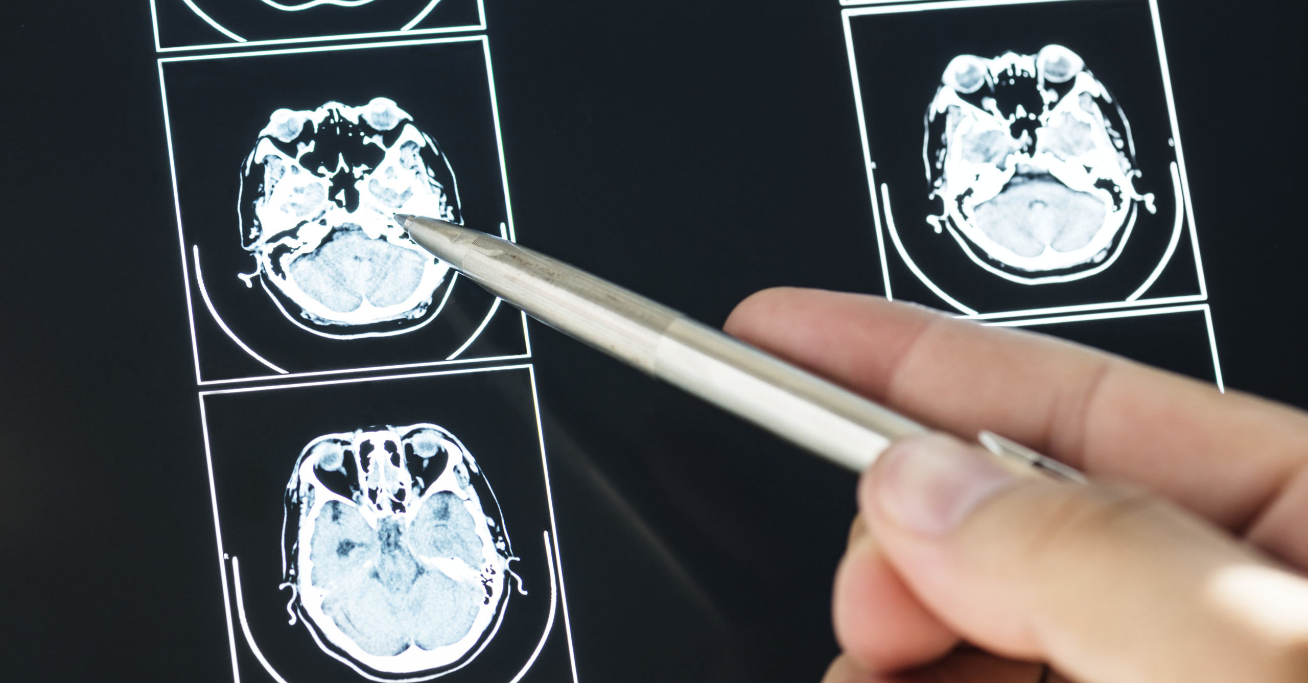
Stroke is the second most common cause of death worldwide, and the extent of ischaemic damage is the determinant factor for treatment. There is a very narrow time window of a few hours for treatment initiation, but point-of-care molecular imaging is lacking.
To address this problem, the EU-funded GAMMA-MRI project is proposing the development of a breakthrough imaging technology manipulating gamma rays that combines MRI with nuclear medicine imaging principles (gamma emitter radioisotopes), combining the imaging advantages of these two technologies. This novel device incorporates artificial intelligence and is expected to advance molecular imaging while bringing the personalised early management of stroke a step closer to reality.
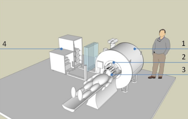
The Gamma-MRI project is dedicated to developing a clinical molecular imaging device based on the physical principle of anisotropic gamma emission from hyperpolarised metastable xenon. In the strategic move from “one size fits all” to personalised medicine, molecular imaging plays an essential role. However, despite significant technological advances in the last decades, medical imaging (especially for the brain) relies heavily on very expensive, complex, and bulky machines. Moreover, MRI suffers from low sensitivity, only partially compensated by the recent advances in hyperpolarisation. On the other hand, the very sensitive PET and SPECT imaging modalities offer limited spatial resolution. Besides those trade-offs, the limited access to suitable devices still hinders the applicability of medical imaging to address major healthcare challenges in brain-related pathologies, even in Europe.
Stroke alone is the second cause of death and the third cause of disability worldwide. The evolution of ischaemic damage varies much among patients. To achieve significant improvement in the outcome of the patients, a careful selection of the treatment path guided by images of the ischaemic brain, in a narrow time window of just a few hours is crucial. Unfortunately, point-of-care molecular imaging that could speed up patient management barely exists. Gamma-MRI is a game-changer imaging technology, combining the high sensitivity of gamma ray detection and the high resolution and flexibility of MRI, bringing down by multiple fold the cost of molecular imaging. Six closely interlinked work packages will cover: production of hyperpolarised gamma-emitting xenon isomers; preserving hyperpolarisation until delivery to targeted organ; developing advanced image acquisition and reconstruction using physics- and artificial intelligence- based approaches; designing and assembling the prototype upon a low field versatile magnet; and implementing the first preclinical Gamma-MRI brain imaging experiment.
Alternatives to Gadolinium
RS2D successfully applied and was selected for a FETOpen project (2019-2022) focusing on Hyperpolarized MR technologies and molecular probes as alternatives for conventional metal-containing contrasts agents. It’s a three-year collaborative research project that brings together highly experienced engineers, physicists, chemists and radiologists to develop a radically new and innovative contrasts agents for magnetic resonance imaging (MRI).
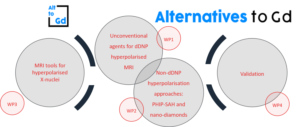
The specific aims AlternativesToGd are:
- Development of unique hyperpolarised agents via dissolution-dynamic nuclear polarisation (dDNP) and administration routes that produce long lasting and intense signal in the blood pool
- Development of hyperpolarised small-molecules for biomedical imaging using parahydrogen-induced hyperpolarisation (PHIP)
- Development of new hyperpolarisation techniques based on optical or DNP irradiation of nano-diamonds
- Development of tailored MRI tools for optimal observation of the hyperpolarised agents in vivo
Although the project aims at finding at least one replacement for the standard Gd contrast agent used today, by exploring a variety of molecules, methods and hyperpolarization techniques, there is a potential to combine these and administer a “cocktail” comprised of two or more hyperpolarised molecules. By looking to a “cocktail solution” there is a potential to optimize the decay time and biodistribution properties to enable in depth characterization of the tissue microenvironments that could provide better results than what is currently achieved with available contrasting agents.
The use of hyperpolarised agents for perfusion imaging or tissue-retention imaging for diagnostic MRI is a new technology that does not exist today. We will lay the foundation for such a technology by providing the contrast agents, the means to increase their MR signal, and the routes for MR imaging of these hyperpolarised signals on clinical MRI scanners. Society will gain by having access to medical imaging that does not leave potentially toxic deposits in the bodies of children and adults undergoing MRI examinations. Our results also have the potential to access new markets for medical imaging and contrast media companies, whether established or young start-ups.
More info on the AlternativesToGd website.
TRIMAGE
In 2013, RS2D succeed to a European Framework Program, FP7 (2013-2017), to build an optimized trimodally (PET/MR/EEG) clinical imaging tool for schizophrenia.

The aim of the project was to design an optimized and a cost-effective trimodally imaging instrument (brain PET/MR/EEG scanner) for early diagnosis, monitoring, and follow-up of schizophrenia disorders. RS2D was the main industrial partner of the project, in charge of the development and construction of the MR part.
The aim of the project was to integrate three well-known imaging modalities in their current clinical applications and to build a new dedicated instrument. In combination with biomarkers from high-end European research laboratories, the new tri-modal PET / MR / EEG scanner is a fully integrated solution for the early diagnosis of schizophrenia.
The consortium develops and build a PET imaging system with better performance than current clinical imaging: spatial resolution of 2 mm and high sensitivity (14%). The PET ring is based on the silicon photomultipliers technology in order to be compatible with the magnetic field and is as compact as possible.
The MRI is based on an ultra-compact superconducting magnet, without the need of cryogenic fluids, and use the latest technology in this field. The electronics developed by RS2D is performing rapid imaging, localized spectroscopy and diffusion weighted imaging.
The PET and the MR is fully integrated in the same compact enclosure and also integrate the electroencephalography system (EEG). This new system is optimized for human brain studies, with improved patient comfort and reduced overall cost compared to existing systems.