NMR and MRI samples
NMR & MRI, what do I do with this sample ?
We at RS2D love to go deep on electronics and the basic principle of NMR and MRI. For users of the coolest analytical techniques out there one crucial component of measurements might be even more interesting. Thus, we hope you’ll find this blog post useful – today we talk everything SAMPLE.
What kind of samples can be analyzed with which instrument? Are there any restrictions, limitations, or requirements? And so many other questions. We know that MRI is a medical imaging technique for analyzing living organisms, while NMR is a spectroscopic technique to study molecular structures. Let’s start with an overview of the samples that can be analyzed in Magnetic Resonance technologies.
Samples
In MRI, we mainly work on living samples, such as humans, animals, tissues, flora or fruit & vegetables. The sample is directly inserted into the probe, see figure 1, or into the patient’s bed support. Consider a preclinical MRI system, the mouse (or any other small sample) is placed into the probe, and sensors (breath and cardiac monitoring) are placed on it. In clinical MRI, the patient is placed on a mobile bed, and the probe is placed on the patient (probes are specific to the area under study, as for example head probe, in the figure 2).
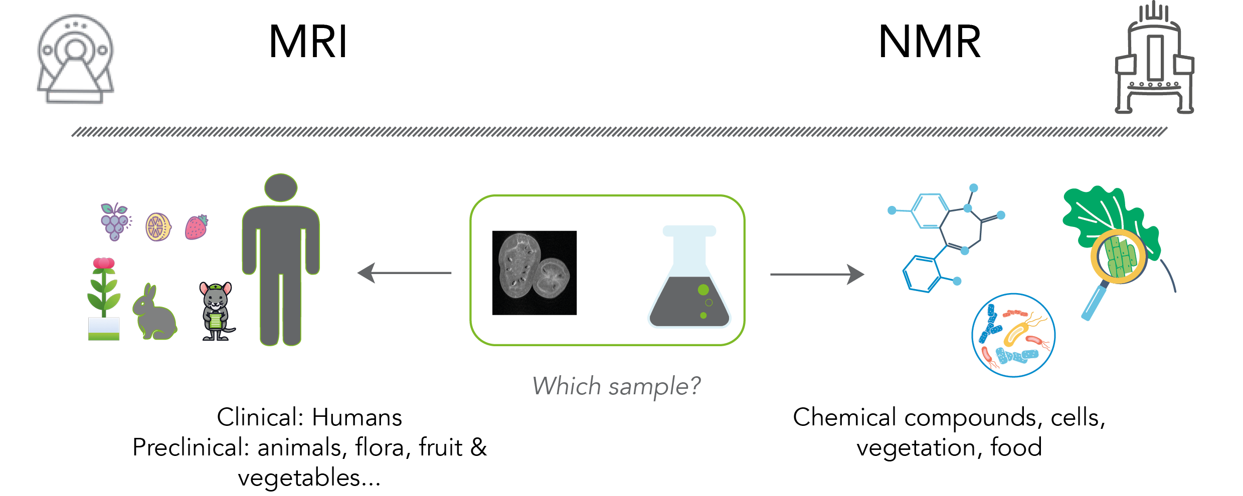
Figure 1: Which sample can be analyzed by NMR & MRI
In NMR, liquid, solid or semi-solid samples can be analyzed. These can be chemical compounds (small or macromolecules), cells, vegetation [1], food [2] or any solid-state or liquid-state sample. The sample is placed in the appropriate vessel: an NMR tube for a liquid sample, a HR-MAS (High Resolution Magic Angle Spinning) rotor for solid or soft samples. A flow cell can be used instead of NMR tubes, for reaction monitoring or hyphenated NMR like LC-NMR. In each case, the sample has to be prepared according its final vessel.
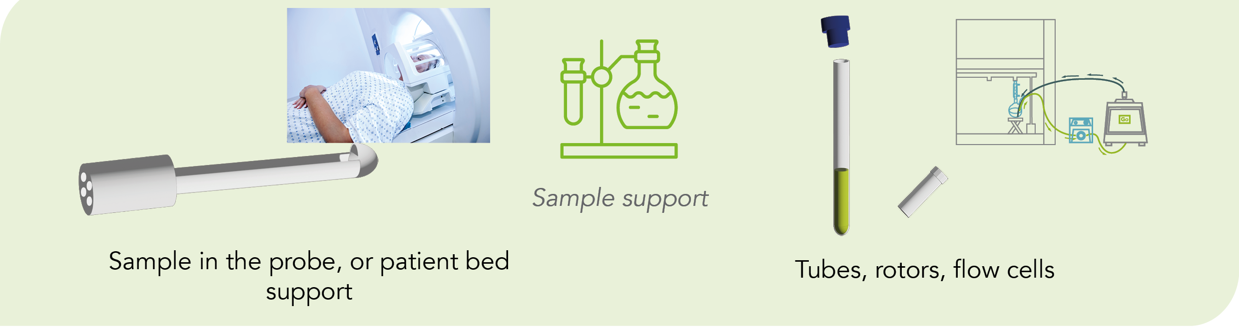
Figure 2: Sample support
Preparation
As magnetic resonance spectroscopy is a non-destructive technique and the preparation is easy. In MRI, the patient or sample should not have any magnetic parts (e.g. metallic implant). To improve the sensitivity of internal structures, a contrast agent [3] can be used, see our Gadolinium project. A contrast agent is a chemical compound injected into the patient to improve contrasts in the image. They are paramagnetic and based on Gadolinium. Recently it has been observed a toxicity when using Gd compounds, so our project is to develop new safe contrasts agents for MRI.
In clinical MRI, patient positioning can be delicate and some rules must be followed [4] to ensure good image quality. In preclinical MRI, when working on living animals, user must put the sample to sleep with an isoflurane anesthetic gas.
In NMR, same rule applies regarding the absence of magnetic parts in the sample. This could damage the NMR spectrometer. NMR experiments are non-destructive, unless the user needs to perform a temperature variation study on a thermosensitive sample. One of the main requirements in liquid NMR is to use a deuterated solvent. In most cases the user will choose a deuterated solvent, but sometimes (large scale reaction monitoring in flow, neat sample measurement) this is not possible and 1H lock will be employed.
In liquid state NMR, it’s important to work with homogenous samples, as NMR experiments require a uniform magnetic field over the whole of the NMR sample volume to get sharp signal. A non-homogenous sample will provide a broad NMR spectra. When working on semi-solid or solid state sample, user may work with the HR-MAS (High Resolution Magic Angle Spinning) NMR technique to remove broadening effect. The sample is packed into a rotor. This will be further detailed in a dedicated article. Stay tunned !
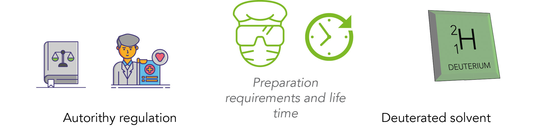
Figure 3: Life time and preparation requirements
Lifetime and regulation
Lifetime of a sample can be minutes to hours depending on its nature. A clinical or preclinical subject will not be able to stay for more than 30 minutes without moving in an MRI system. When working on a non-living sample, it may be easier to keep the sample stable for hours due to the temperature regulation in the spectrometers. In NMR, there are no specific regulations about samples, unlike MRI where Authority Regulations are required to work on living samples, figure 3. Guidelines for the protection of animals used for scientific purposed are established and must be strictly followed [5].
Physico-chemical properties
NMR and MRI systems allows user to monitor and control some physico-chemical parameters. When working in NMR, the temperature must be controlled and regulated. However other parameters are important and must be handled carefully before doing NMR analysis, as the pH. The salinity of the sample has an impact the chemical shift of the line width. The sample has to be stable during the experiment, and should not deteriorate, as this will be observed on the final data.
In MRI, more parameters are monitored and managed by the system. When working with a living sample, temperature must be regulated by heating the bed, respiration and heart rate must be monitored.
Dimensions
Both NMR and MRI allows the user to work in several dimensions. In MRI, we can work in 2D or 3D and the planes are well defined by: Axial, Coronal and Sagittal planes. See figure 4. NMR allows user to perform acquisitions in 2D, 3D and even more when working on macromolecules [6].
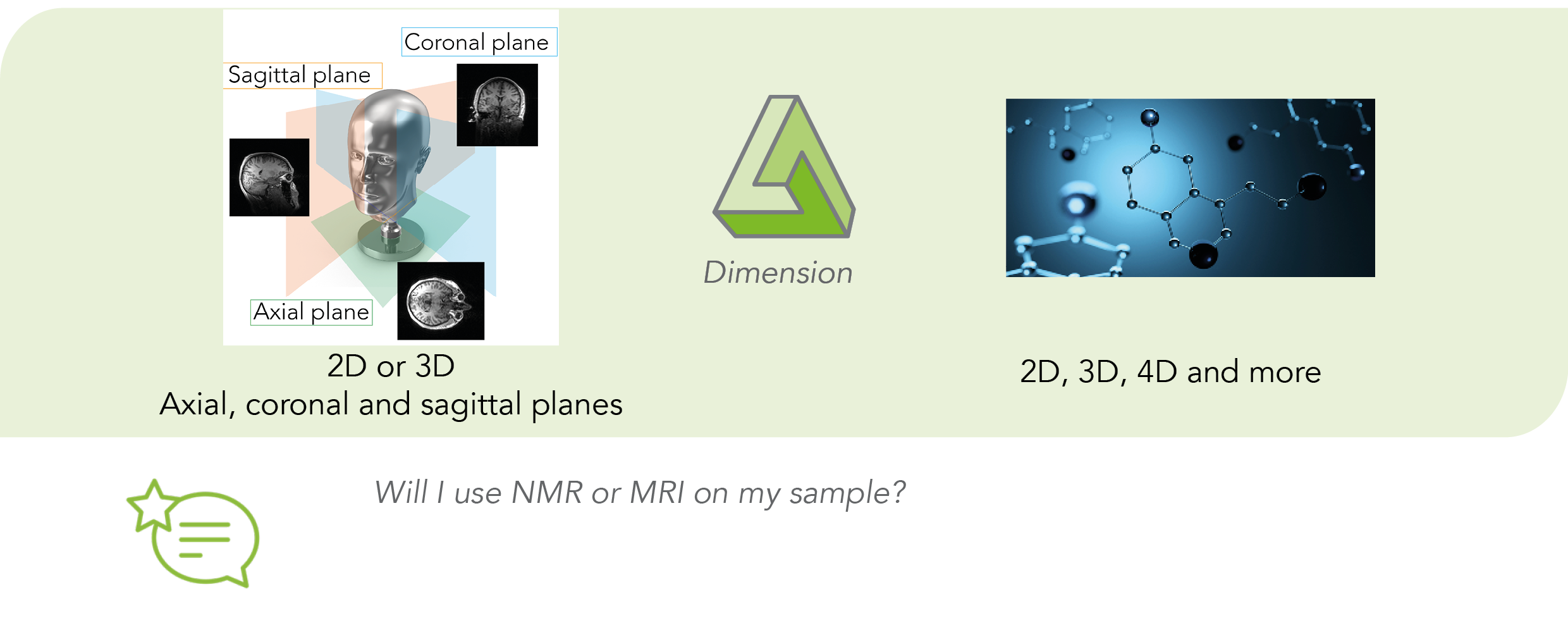
Figure 4: Dimensions and planes in MRI and NMR
Summary
Now, consider a lemon analysis. If you need to observe the whole lemon, you will have to use MRI, to observe density proton location into the lemon, and obtain an image of the inside of the lemon. But if you want to see the proton presence and the molecular structure of you lemon, you will have to do NMR.
If you have any questions regarding sample preparation or anything else about NMR and MRI, please contact us.
Bibliography
[1] Targeted discovery of bioactive natural products using a pharmacophoric deconvolution strategy: Proof of principle with eleganolone from bifurcaria bifurcata R. Ross. F. Nardella and al, M. Bourjot. Phytochemistry Letters 26 (2018) 138–142
[2] L. Shintu, S. Caldarelli, High-resolution MAS NMR and chemometrics: characterization of the ripening of Parmigiano Reggiano cheese, J. Agric. Food Chem. 53 (2005) 4026–4031.
[3] Mendonça-Dias MH, Gaggelli E, Lauterbur PC. Paramagnetic contrast agents in nuclear magnetic resonance medical imaging. Semin Nucl Med. 1983 Oct;13(4):364-76.
[4] https://www.ncbi.nlm.nih.gov/books/NBK565882/
[5] https://eur-lex.europa.eu/LexUriServ/LexUriServ.do?uri=OJ:L:2010:276:0033:0079:fr:PDF
[6] 4D solid-state NMR for protein structure determination. Phys. Chem. Chem. Phys., 2012,14, 5239-5246