
A Total Eclipse of the Heart through MRI
Can you feel the love in the air? If you’re lucky, your Valentine’s Day may be met with heavy palpitations, sweaty palms, and general nervousness. Although you can feel all this happening throughout your body, what does it really look like internally? In this blog, we will be examining an MRI image of a real heart and if you stay tuned, you might just see it beating!
As February approaches and love begins to fill the air, we all understand what time it is. You guessed it, it’s almost Valentine’s Day, so let’s speak from the heart. Have you ever wondered if the butterflies you feel are something happening physiologically? Let me start by explaining the signs you may feel when you become interested in someone. You may feel your heart rate increasing, nervousness taking over and you may even begin to blush, all telltale signs show
ing you that there may be something deeper. The question then becomes, “Do I feel an attraction?” Well, maybe, but let’s see for ourselves through the use of Magnetic Resonance Imaging (MRI). Yes, you heard it, we will be taking an MRI of a real heart!
You may be thinking, why don’t we use an X-ray or CT scan? Well, MRI can tell us a different story than a CT or X-ray can. When two people fall in love, changes occur in the body. Hormones, such as oxytocin, endorphins, dopamine, and serotonin, can give you the feeling of happiness, making your heart beat louder and quicker. With traditional imaging techniques it can be difficult to obtain the image of a heart as it is always moving (beating); however, scientists have developed a particular MRI experiment that can capture the motion of a moving organ. This experiment is called a “Cine cardiac” MRI sequence.1 A cine cardiac sequence is a repetition of images of the same area at the same rhythm. This experiment relies on the use of an electrocardiogram (ECG) signal to synchronize the acquisition of imaging data with cardiac motion.2 As this experiment requires a fast-imaging technique, our Cameleon4TM MR electronic platform is the perfect fit for this type of study.
As an example, let’s discuss the following clinical image of a chest cavity (Figure 1), which was acquired using a Nanoscan 3T MRI system.
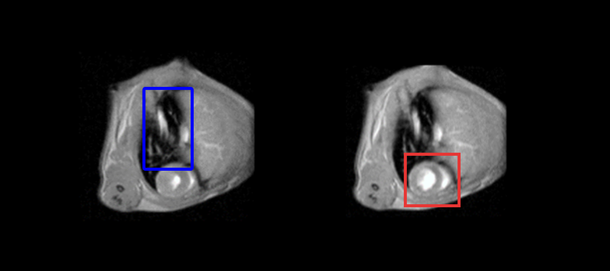
Figure 1: MRI image of a chest cavity area using Nanoscan 3T MRI system. Left ventricular axis image of a chest cavity, where the lung is outlined in blue and heart outlined in red. Within those organs, the black, white, and grey portions represent the air, blood, and muscle, respectively.
Upon first glance, you may wonder, “What am I even looking at?” MRI images are defined using a black and white scale, which ultimately represents the density of hydrogen atoms. That being said, MRI is an imaging technique that studies the magnetic properties of hydrogen nuclei. Although they only represent 10% of the body’s mass, hydrogen atoms account for 86% of our chemical composition, which are mainly distributed throughout the water and fat molecules of the body.3 This means, in MRI we can distinguish between fat and water through the density of hydrogen. Ultimately, that density is referred to as the signal and is measured through a gray scale. Depending on the type of MRI experiment used (ie. T1- or T2- weighted), the corresponding image is represented in a grayscale shown in Table 1.
| T1-weighted | T2-weighted | |
|---|---|---|
| Air, compact bone | Black | Black |
| Water | Black-grey | White |
| Tissue | Grey | Grey |
| Fat | White | Light-grey |
Table 1: T1- and T2- weighted experiments greyscale, where these experiments are played by the radiological manipulator according to the doctor’s specification.
If we refer back to Figure 1, within the cavity, we can now see that the blood appears in white and is concentrated in the heart, whereas the air in the chest cavity (specifically the lungs) appears in black.
To determine if your heart is beating well and if your blood flow is regular, we combined a series of MRI images into a motion picture to observe the heart’s movement and blood flow, which is shown in Figure 2.
 |
 |
Figure 2: Animated GIF of the chest cavity studied by MRI where the left GIF shows a heart beat of 60 beats per minute (bpm) and right side of 160 bpm.
Now that we can check if our heart is working properly, let’s go back to our original question: do I feel an attraction? Well, if your heart isn’t beating very fast, similar to the left GIF of Figure 2, then probably not. On the other hand, if your palms are sweaty, there are butterflies in your stomach, and your heart is similar to the right GIF of Figure 2, then it might just be love (or a serious exercise). For all you single people out there, let’s look on the bright side, you can now show off your newfound ability in reading an MRI image!
Bibliography
[1] Atkinson, D. J.; Edelman, R. R. Radiology 1991, 178, 357-360.
[2] Rokey, R.; Wendt, R. E.; Johnston, D. L. Magn. Reson. Med. 1988, 6(2), 240-245.
[3] https://www.news-medical.net/life-sciences/What-Chemical-Elements-are-Found-in-the-Human-Body.aspx (accessed 2022-08)
