Magnetic Fields in Magnetic Resonance Imaging (MRI)
I read the most insane article today: “Lawyer dies after shot by his own concealed gun triggered by MRI scanner[1]” and thought it’s a good time to discuss the aspects of strong magnetic fields used in MRI, and MRI safety.
For those who have yet to experience an MRI scan, you may not know that to generate these powerful diagnostic images, you must be placed directly inside the magnet. Most MRI spectrometers use superconducting magnets, some use permanent magnets, but regardless of how the magnetic field is generated, the requirement is still the same, the ‘sample’ must be placed into the magnet.
So, let’s discuss what this means, practically. There are magnetic vectors that go from the south pole of a magnet to the north pole of the magnet (figure 1), and these magnetic vectors are not completely contained inside the material, but rather extend outside in what’s called a ‘stray-field’, ‘fringe-field’ or ‘demagnetizing field’. You may have seen this with your own eyes if you remember elementary school science experiments with magnets and iron filings. These magnetic fields extend outside the magnet and get weaker and weaker as you move further away.
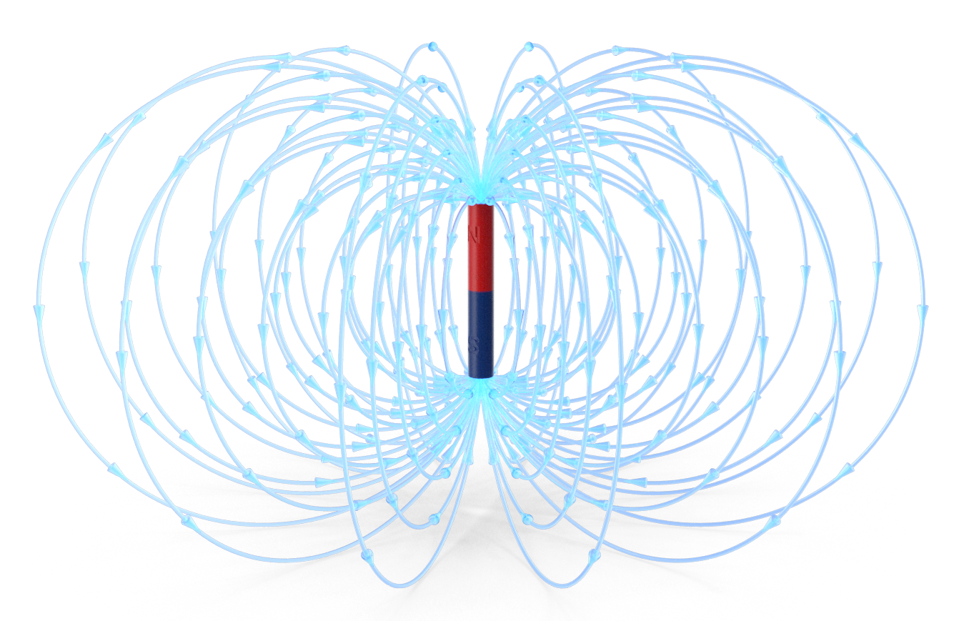
Figure 1: A magnet with the North pole shown in red and the South pole shown in blue. Magnetic field lines are light blue and go from South to North.
A material containing a magnet component, such as a bullet, if too close to the magnetic field may find itself magnetically attracted to the field, and the closer it gets, the stronger this force of attraction becomes, until eventually this force dominates, and the magnetic material gets drawn into the field.
To illustrate the extent of these magnetic forces when you enter a room containing an MRI (or NMR, for that matter), you may notice that there are lines marked on the floor. These reveal the distance from the magnet where the stray field is 5 Gauss (i.e., 10x greater than the earth’s magnetic field), and the area where potentially magnetic material, like the bullet, start to be attracted to the magnet. The distance of these lines from the magnet depends on the strength of the magnetic field, the magnet design/configuration (e.g., superconducting vs. pole and yoke vs. Halbach configuration) and the magnetic shielding used to protect the instrument.[2]
To help illustrate[3], an unshielded 1.5 T MRI, a field greater than 0.5 mT (5 Gauss) extends to about 12 meters from the magnet center and 9.5 meters on either side of the magnet! A 3T nanoscan MRI preclinical system, however, which is actively shielded, has a residual field strength below 0.8 m on side of the magnet.
Sidebar: To interconvert the two units often used to discuss magnetic fields: 1 Tesla [T] = 10 000 Gauss [G].
While safer to those outside the MRI, it is important to note that active shielding does make the magnetic field more intense at the entrance of the spectrometer. So regardless if the instrument is shielded or not, these lines should be respected as is illustrated in this alarming video: https://www.youtube.com/watch?v=xn6sDYOrOC8.
In our next blog, we will discuss different types of magnetic designs and configurations, starting with cryocooling part the magnet. If you have questions or would like to learn more about the RS2D MRI product line please don’t hesitate to contact us!
References
[3] https://www.techno-science.net/glossaire-definition/Imagerie-par-resonance-magnetique-page-3.html