
What is PET-MRI ?
Among the preclinical imaging material RS2D offers, one can find the PET-MRI system. Then, what is PET-MRI? Following the first blog laying the foundations of nuclear magnetic resonance in MRI and NMR, let’s discuss about this combined system. This article will be focus on MRI hyphenated technique: PET-MRI imaging. For further NMR hyphenated technique, follow us to stay updated.
Positron Emission Tomography, PET, is a non-invasive nuclear medicine imaging technique (clinical and preclinical) to measure in three dimensions the metabolic or molecular activity of an organ. The system is like an MRI spectrometer: a circle with hole where the sample is inserted. Let’s first present the PET system, commonly named PET scan.
Concept
PET uses molecules labeled with positron emitting isotopes as molecular probe for quantification of in vivo radioactivity through the emissions produced by an intravenously injected product.
This product called a tracer is a radioactive isotope compound and is selected according to the targeted tissue or organ.
There is an increasing list of these tracer which are being used for PET imaging [1]. The list of some of commonly used radioactive isotopes compounds is (radioactive isotope in parentheses):
- Fluorodeoxyglucose, FDG, most common PET radiotracer (F-18)
- Sodium fluoride, fluoro-ethyl-spiperone (F-18)
- Acetate, Choline or Methionine (C-11)
- Prostate-Specific Membrane Antigen, PSMA (Ga-68)
- Rubidium chloride (Rb-82)
- Ammonia (N-13)
PET scan is used in medicine to detect cancers by measuring the cellular activity. It detects tumors, their severity and size, and following the effectiveness of treatments and identification of possible metastases. This technique is less precise in comparison to an MRI or scanner, that’s why PET is often used in combination with MRI or CT to get a detailed image.
PET signal is obtained thanks to the tracer, which produces a particle catch by a detector present in the ring of the PET machine.
Material
Here is a schematic view of a PET ring, with the scintillator crystals and detectors, and a schematic view of our PET-MRI 3T Nanoscan product.
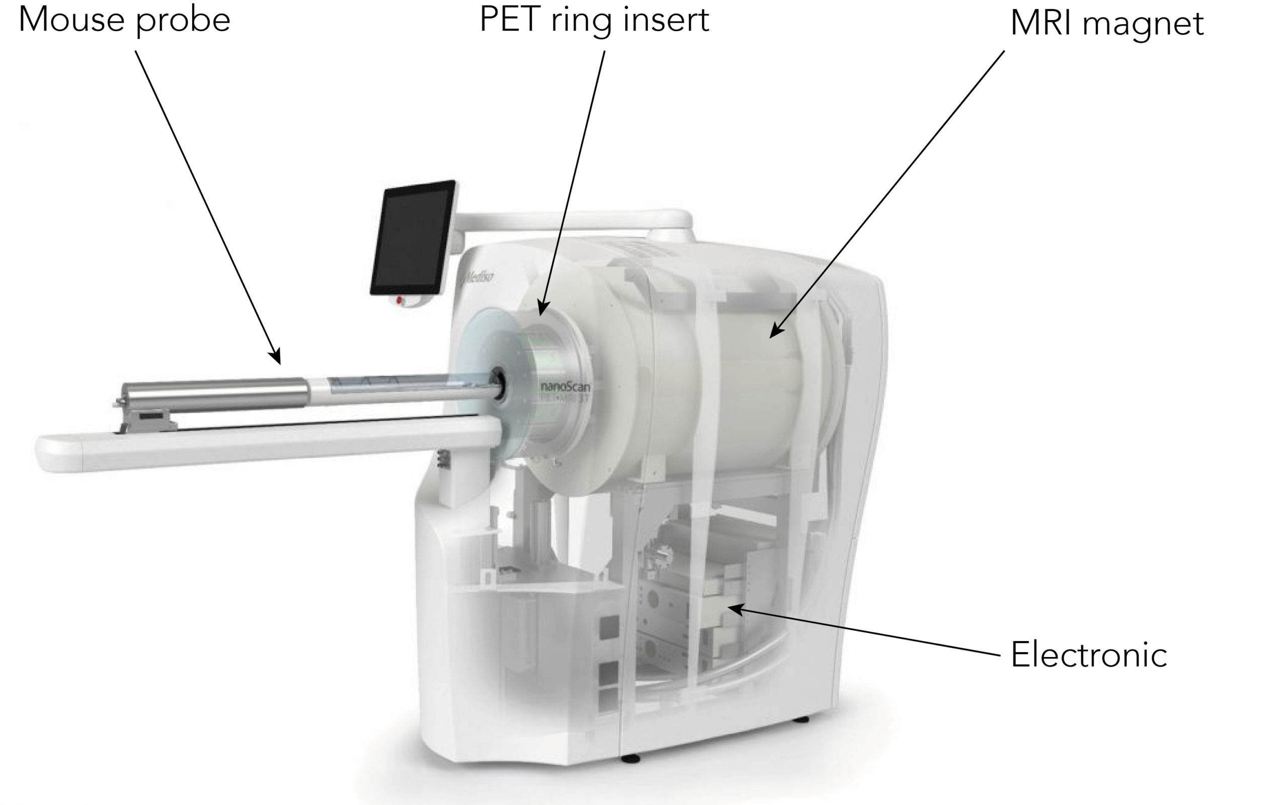
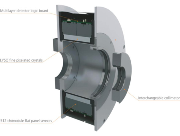
Figure 1, left: Inside view of a PET-MRI system
Figure 2, right: PET ring description
PET-MRI results
PET data acquisition needs to be synchronized with breathing movement and heartbeats as they are performed in preclinical or clinical imaging. PET data can be reconstructed and displayed as 3D images, and the physiologic information afforded is priceless, the quality of image is poor, noisy with a limited spatial resolution. That’s why this technique is often combined with CT or MRI spectroscopy.
Here are some data example, provided by Mediso.
Multiple animal imaging with PET/MRI 3T
Simultaneous scan of 3 tumor-bearing mice
Animal model: BALB/c mice
Radiotracer: 18F-FDG, 4.87 MBq (131.6 μCi), 4.75 MBq (128.3 μCi) and 5.91 MBq (159.7 μCi)
Acquisition: GRE 3D Multi-FOV MRI, acq. time: 18min, NEX: 4, TR: 10ms, TE: 3.1ms, TH: 0.8mm, PET acquisition: static
RF Coil: 72mm Quadrature Tx/Rx volume coil
18F-FDG Glioma imaging in mouse brain
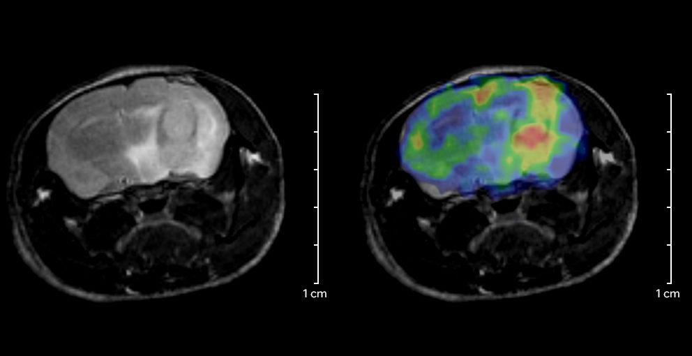
MRI is currently the gold-standard in glioma imaging, thanks to its capabilities in delineating structures within soft tissues. Combining with the molecular specificity of PET, the nanoScan® PET/MRI spectrometers are the perfect tool for the development of novel therapeutic and diagnostic strategies for glioma.
Animal model: C56BL/6 mouse (28g)
Tracer: 3.2 MBq (86 µCi) 18F-FDG
Acquisition: T2W FSE 2D, FOV: 32mm x 32mm, TH: 1mm, MRI acq time: ∼5 min, PET acq: dynamic
Coils: Quadrature Tx/Rx volume coil for mouse brain
You would like to know more details about PET, PET-MRI or preclinical imaging systems, contact us anytime you need.
You wish to know more about PET principle? Stay tuned on our twitter to read it soon.
Bibliography
[1] https://radiopaedia.org/articles/positron-emission-tomography